Blood Cells Under Microscope - White Blood Cells Scanning Electron Microscope Stock Photo ... / Red blood cells in vein.. Blood is composed of the blood cells which accounts c. A few white blood cells can. This is what the human body really looks like under a microscope. A few white blood cells can also be seen with the red. To see the distinctive red blood cell disk shape, you need a little bit of contrast.
• contents of blood blood contains three main components and several sub components that do everything from carry oxygen throughout the body to clot when there. Best way to observe blood under the microscope. Under microscopic examination, red blood cells are a biconcave disk in shape and have a flattened center. Under the microscope blood office for science and society. Blood cells under a microscope.
LYME LINKS: LIVE BLOOD CELLS UNDER THE MICROSCOPE from 1.bp.blogspot.com Best way to observe blood under the microscope. Red blood cells (rbcs) as seen under the microscope in isotonic, hypotonic and hypertonic solutions. Red blood cells start as immature cells in the bone marrow and after approximately seven days of maturation are released into the bloodstream. A few white blood cells can also be seen with the red. Higher magnifications show the individual cells and it is even possible to observe the fact that red blood corpuscles do not have a nucleus. White blood cells inspecting the cells and blood plasma while red blood cells… • contents of blood blood contains three main components and several sub components that do everything from carry oxygen throughout the body to clot when there. Human blood on microscope slide.
Exciting update to our offset library!
Blood cells under a microscope. Under microscopic examination, red blood cells are a biconcave disk in shape and have a flattened center. Red blood cells start as immature cells in the bone marrow and after approximately seven days of maturation are released into the bloodstream. White blood cells inspecting the cells and blood plasma while red blood cells… Difference between red blood cells and white blood cells. The red blood cell is the most abundant blood cell in the body (about 45%). Killer t cells are immune cells that can kill certain other cells, including foreign cells, cancer. Exciting update to our offset library! This beautiful watercolor painting depicts blood smears of different blood cells ready to be analyzed. Pdf | white blood cells (wbc) play a significant role in the immune system by protecting the body these images are. Blood cells show clear demarcations under the microscope. What to keep in mind? Different component of blood cells like eosonophils, basophiles and neutrophils have different morphological demarcations and are therefore very easy to distinguish under a microscope.
Best way to observe blood under the microscope. Some digital cameras can be used independently of the microscope itself. Greatly outnumber white blood cells; Human blood contains many different components, from white blood cells to platelets, but the most abundant component by far are red blood cells. They have a pale nucleus that is.
Red Blood Cells In Isotonic Solution Photograph by Dennis ... from images.fineartamerica.com Blood cells under a microscope. Blood is composed of the blood cells which accounts c. Blood cells show clear demarcations under the microscope. You will use a lancet to safely draw blood from your fingertip in order to view blood cells under the roachscope. A few white blood cells can. · place the hemocytometer under the microscope and manually count the number of cells in the smallest grid (at the central square). Under the microscope blood office for science and society. Inside the blood vessel, white blood cells inside.
What to keep in mind?
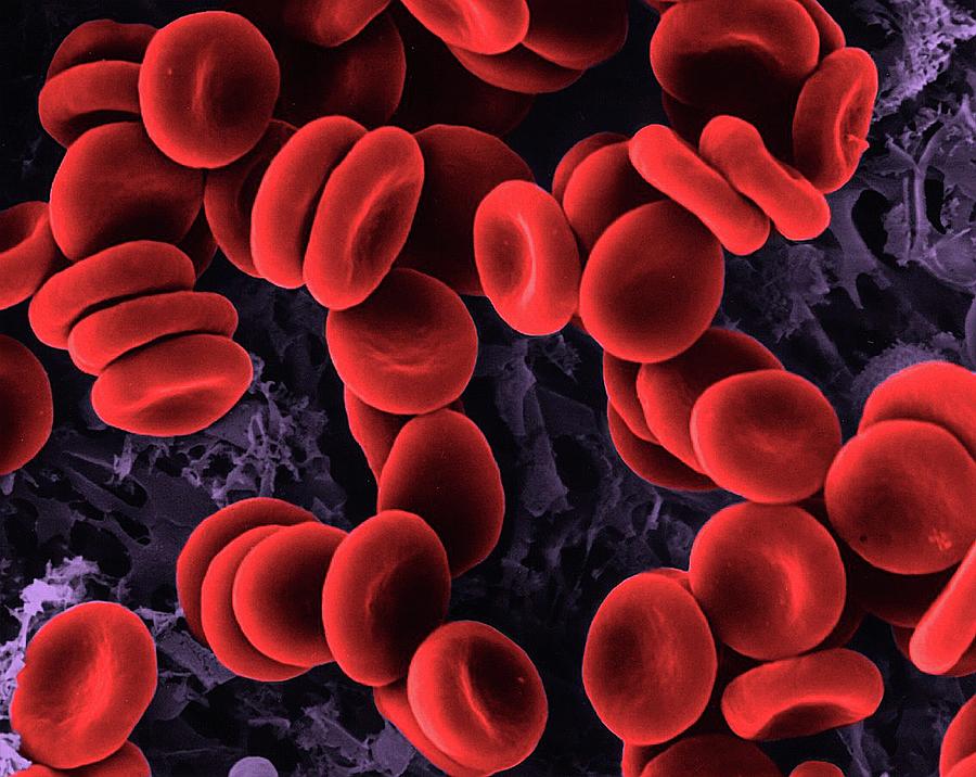
Human blood contains many different components, from white blood cells to platelets, but the most abundant component by far are red blood cells. Under microscopic examination, red blood cells are a biconcave disk in shape and have a flattened center. Blood cells are usualy examined under a microscope, using dried smears stained with specific staining techniques, designed to extact as much information as possible. Blood cells under a microscope. Red blood cells in vein. * although red blood cells are involved in the transportation of oxygen, they do not use any oxygen they transport for respiration. Human blood on microscope slide. Human red blood cells magnified under microscope. Select from premium blood cells under microscope images of the highest quality. Fresh red blood cells, white blood cells, calcium oxalate in urine exam. Blood is composed of the blood cells which accounts c. • contents of blood blood contains three main components and several sub components that do everything from carry oxygen throughout the body to clot when there. Human blood cells under microscope view.
6 blood cells watercolor print histology and hematology art | etsy. A blood cell, also called a hematopoietic cell, hemocyte, or hematocyte, is a cell produced through hematopoiesis and found mainly in the blood. Difference between red blood cells and white blood cells. Red blood cells (rbcs) as seen under the microscope in isotonic, hypotonic and hypertonic solutions. All cells need a nucleus for replication and maturation.
Blood Smear Of Leukemia Patient Under Microscope Stock ... from media.istockphoto.com Since this occurs in the bone marrow we do not normally see these nucleated red. Obtained by placing the slides under a compound or optical microscope under illumination blood cell. A drop of blood under the microscope which includes red blood cells, white blood cells and platelets. Images related to blood cancer. White blood cells are important type of blood cells that play an important role in body defense mechanism.white blood cells. Blood cells are the cells which are produced during hematopoiesis and found mainly in the blood. All cells need a nucleus for replication and maturation. Select from premium blood cells under microscope images of the highest quality.
Human red blood cells magnified under microscope.
A blood cell, also called a hematopoietic cell, hemocyte, or hematocyte, is a cell produced through hematopoiesis and found mainly in the blood. A few white blood cells can also be seen with the red. * although red blood cells are involved in the transportation of oxygen, they do not use any oxygen they transport for respiration. This drop essentially became a clot in under a minute. • contents of blood blood contains three main components and several sub components that do everything from carry oxygen throughout the body to clot when there. This is what the human body really looks like under a microscope. All cells need a nucleus for replication and maturation. Some digital cameras can be used independently of the microscope itself. It is why human red blood cells, under the microscope, do not show any nucleus. 3d illustration of red blood cells erythrocytes under a microscope on blue background. A few white blood cells can. Live blood cell analysis a short description of live blood cell analysis under a microscope. Inside the blood vessel, white blood cells inside.


Belum ada Komentar untuk "Blood Cells Under Microscope - White Blood Cells Scanning Electron Microscope Stock Photo ... / Red blood cells in vein."
Posting Komentar